Lecture Details[]
Colin McHenry; Week 10 MED1022; Anatomy
Lecture Content[]
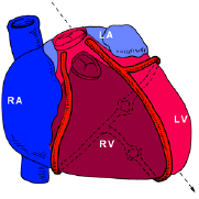
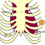
Pericardial sac has visceral (serous) and parietal (serous/fibrous) parts, has reflections for vessels, sinuses, pericardial cavity, heart, ascending aorta, pulmonary trunk, lower half SVC, termination of IVC, termination of pulmonary veins. Heart is unpaired viscus of 2 halves, 1/3 to the right. Apex is the left ventricle, anterior at 5th IC space, base is left atrium and is posterior. Right border is right atrium, upper border is 3rd to 6th CC, curved left border is left ventricle, apex to lower border at 2nd costal cartilage, other borders are two straight lines.
There is a sternocostal (right ventricle) and diaphragmatic (both ventricles) surface. Walls are myocardium lined by endocardium covered by epicardium. Right atrium is smooth, rough
anteriorally, has crista terminalis, right auricle, right AV orifice, interatrial septum. Right ventricle has papillary muscles, trabeculae carnae, moderator band to anterior papillary muscle (contains right bundle branch), conus arteriosus (conical pouch) where the pulmonary trunk arises. Left ventricle has trabeculae carnae, mitral valve, IV septum, aortic vestibule, aortic orifice, valve (left, right and posterior cups). Cardiac skeleton is made of fibrous rings surrounding the four valves, forms an electrical barrier with a gap for the conducting system. Sinoatrial node is at the top of sulcus terminalis, anterior to SVC, AV node is in IV septum within Koch's triangle, AV bundle of His in the membranous septum. Right and left bundle

branches are in the muscular septum surrounded by the moderator band at the right. From the aorta there are right and left coronary branches with potential anastomoses. RCA is to the right of the pulmonary trunk in the right AV groove, terminates after giving off posterior IV branch. LCA is to the left of the pulmonary trunk, divides into anterior IV and circumflex branches, circumflex runs in left AV groove. RCA is usually dominant. SA nodal branch is usualy from RCA (55%), AV nodal branch usually from right (90%).
Coronary sinus is in AV groove posteriorly, opens into the right atrium. There are great, middle and small cardiac veins which run with the arteries. Anterior cardiac veins drain into the right atrium. Venae cordis minimae drain directly into chambers. Oblique vein of the left atrium is remnant of left SVC and may persist.
Myocardial pain is in chest and inner arm (T1), can have atypical pain. Pericardial pain is to the shoulder tip (C4). Pleuritic pain has costal pleura to chest, mediastinal to shoulder tip. Diaphragmatic pleura has central pain to shoulder tip, peripheral to anterior abdominal wall (7-12).